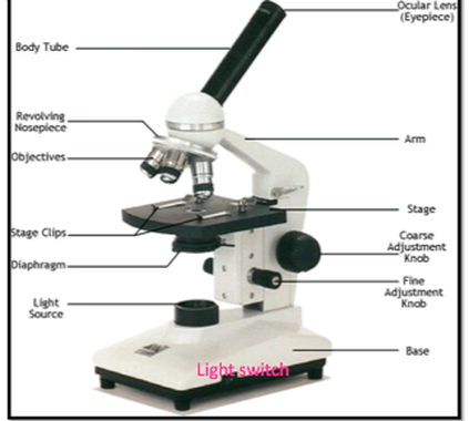
38 light microscope with labels
Simple Microscope - Diagram (Parts labelled), Principle, Formula and Uses A simple microscope consists of Optical parts Mechanical parts Labeled Diagram of simple microscope parts Optical parts The optical parts of a simple microscope include Lens Mirror Eyepiece Lens A simple microscope uses biconvex lens to magnify the image of a specimen under focus. Label the microscope — Science Learning Hub 08/06/2018 · All microscopes share features in common. In this interactive, you can label the different parts of a microscope. Use this with the Microscope parts activity to help students identify and label the main parts of a microscope and then describe their functions.. Drag and drop the text labels onto the microscope diagram. If you want to redo an answer, click on the …
Compound Microscope Parts - Labeled Diagram and their Functions The eyepiece (or ocular lens) is the lens part at the top of a microscope that the viewer looks through. The standard eyepiece has a magnification of 10x. You may exchange with an optional eyepiece ranging from 5x - 30x. [In this figure] The structure inside an eyepiece. The current design of the eyepiece is no longer a single convex lens.

Light microscope with labels
Light Microscope- Definition, Principle, Types, Parts, Labeled Diagram ... Apr 07, 2022 · A light microscope is a biology laboratory instrument or tool, that uses visible light to detect and magnify very small objects and enlarge them. They use lenses to focus light on the specimen, magnifying it thus producing an image. The specimen is normally placed close to the microscopic lens. Labeling the Parts of the Microscope | Microscope activity, Science ... Description Worksheet identifying the parts of the compound light microscope. Answer key: 1. Body tube 2. Revolving nosepiece 3. Low power objective 4. Medium power objective 5. High power objective 6. Stage clips 7. Diaphragm 8. Light source 9. Eyepiece 10. Arm 11. Stage 12. Coarse adjustment knob 13. Fine adjustment knob 14. Base Microscope Drawing And Label - Painting Valley All the best Microscope Drawing And Label 33+ collected on this page. Feel free to explore, study and enjoy paintings with PaintingValley.com. ... Compound Light Microscope Drawing. Simple Microscope Drawing. Microscope Drawing. Microscope Drawing Circles. Philippine Map Drawing With Label.
Light microscope with labels. Light Microscope Worksheet Live worksheets > English > Science > Lab equipment > Light Microscope Worksheet. Light Microscope Worksheet. Drag and drop worksheet on the parts of the microscope. ID: 1605909. Language: English. School subject: Science. Grade/level: Middle School. Age: 9-13. Main content: Lab equipment. Label the microscope — Science Learning Hub All microscopes share features in common. In this interactive, you can label the different parts of a microscope. Use this with the Microscope parts activity to help students identify and label the main parts of a microscope and then describe their functions. Drag and drop the text labels onto the microscope diagram. A quick guide to light microscopy in cell biology - PMC Laser-scanning confocal microscopy is excellent for rejecting out-of-focus light and acquiring 3D image data, but in general it is less sensitive and more phototoxic than spinning-disk confocal microscopy. For live specimens for which 3D information is required, spinning-disk confocal microscopy should be considered. Microscope Labeling - The Biology Corner Students label the parts of the microscope in this photo of a basic laboratory light microscope. Can be used for practice or as a quiz. Name_____ Microscope Labeling . Microscope Use: 15. When focusing a specimen, you should always start with the _____ objective.
Microscope Labeling - The Biology Corner May 31, 2018 · Microscope Labeling. This simple worksheet pairs with a lesson on the light microscope, where beginning biology students learn the parts of the light microscope and the steps needed to focus a slide under high power. The labeling worksheet could be used as a quiz or as part of direct instruction where students label the microscope as you go ... Microscope labels Flashcards | Quizlet Microscope label Terms in this set (14) ocular lens / eyepiece diopter adjustment Arm Coarse focus Fine focus On/off switch Base light source iris diaphragm Condenser Stage slide holder Objective lens Nose piece Other sets by this creator Terminology 2 10 terms Laurxyala Centrifuge Skills Test 14 terms Laurxyala Abbreviation 1 10 terms Laurxyala Light Microscope - an overview | ScienceDirect Topics The light microscope is an instrument for visualizing fine detail of an object. It does this by creating a magnified image through the use of a series of glass lenses, which first focus a beam of light onto or through an object, and convex objective lenses to enlarge the image formed. Microscope, Microscope Parts, Labeled Diagram, and Functions Revolving Nosepiece or Turret: Turret is the part of the microscope that holds two or multiple objective lenses and helps to rotate objective lenses and also helps to easily change power. Objective Lenses: Three are 3 or 4 objective lenses on a microscope. The objective lenses almost always consist of 4x, 10x, 40x and 100x powers. The most common eyepiece lens is 10x and when it coupled with ...
What Is PA+++ Sunscreen? [What the Symbol Means] – … 30/10/2017 · When it comes to sun safety, knowledge is power. Skin cancer statistics and sun safety awareness are frightening, and the scary rise in the rates of melanoma has many of us stocking up on the sunscreen—and rightfully so! If you’re looking for ways to keep your skin radiant and safe from the sun’s UV rays, give your sunscreen selection the due diligence it … Microscope Parts and Functions Microscope Parts and Functions With Labeled Diagram and Functions How does a Compound Microscope Work?. Before exploring microscope parts and functions, you should probably understand that the compound light microscope is more complicated than just a microscope with more than one lens.. First, the purpose of a microscope is to magnify a small object or to magnify the fine details of a larger ... Transport of intensity diffraction tomography with non-interferometric ... We present a new label-free three-dimensional (3D) microscopy technique, termed transport of intensity diffraction tomography with non-interferometric synthetic aperture (TIDT-NSA). Without ... Parts of Stereo Microscope (Dissecting microscope) - labeled diagram ... Labeled part diagram of a stereo microscope Major structural parts of a stereo microscope. There are three major structural parts of a stereo microscope. The viewing Head includes the upper part of the microscope, which houses the most critical optical components, including the eyepiece, objective lens, and light source of the microscope.
MINFLUX | Abberior Instruments MINFLUX defines an entirely new class of superresolution methods that uses the best of STED microscopy and the single molecule localization family: 1) Emitters are activated one-at-a-time to obtain the best molecule separation possible, 2) Localization is performed with a light distribution for fluorescence excitation that has a central intensity zero instead of a maximum. This …
The Eye - Science Quiz - GeoGuessr The Eye - Science Quiz: Our eyes are highly specialized organs that take in the light reflected off our surroundings and transform it into electrical impulses to send to the brain. The anatomy of the eye is fascinating, and this quiz game will help you memorize the 12 parts of the eye with ease. Light enters our eyes through the pupil, then passes through a lens and the fluid-filled vitreous ...
Parts of the Microscope with Labeling (also Free Printouts) Parts of the Microscope with Labeling (also Free Printouts) A microscope is one of the invaluable tools in the laboratory setting. It is used to observe things that cannot be seen by the naked eye. Table of Contents 1. Eyepiece 2. Body tube/Head 3. Turret/Nose piece 4. Objective lenses 5. Knobs (fine and coarse) 6. Stage and stage clips 7. Aperture
Animal Cell Under Light Microscope Labelled : Draw and label the ... Most cells are visible under a light microscope, but mitochondria and bacteria are barely visible. Record the microscope images using labelled diagrams or produce digital images. We say cells are microscopic because they can only be seen under a microscope. Note the whiplike flagellum that gives the cell a threadlike appearance.
Barcode Labels and Tags | Zebra 8000T Light-Weight Tag: Tyvek® Olefin Tag: Thermal Transfer: Light-weight olefin label that provides tear resistance and durability. Ideal for lawn tags and garment tags. 8000T Light-Weight Tag. 6000 Wax; 6100 Wax/Resin. 8000T Tuff Tag: V-Max® Polyolefin Tag: Thermal Transfer: Provides good cross tear resistance and outdoor use for up to 1 ...
What is Electron Microscopy? - UMASS Medical School The transmission electron microscope is used to view thin specimens (tissue sections, molecules, etc) through which electrons can pass generating a projection image. The TEM is analogous in many ways to the conventional (compound) light microscope. TEM is used, among other things, to image the interior of cells (in thin sections), the structure of protein molecules …
ZEISS Axiocam Microscope Cameras for Science and Research Discover the monochrome camera for documentation of multiple fluorescence labels in your routine lab. Learn more Axiocam 208 color ... 2.3 Megapixel Microscope Camera: Fast Low Light and Live Cell Imaging High-performance sCMOS microscope camera with a sensor size of 1/1.2" – low read noise for high sensitivity. Learn more Axiocam 705 mono Your fast 5 …
Label the Light Microscope - Labelled diagram - Wordwall Eyepiece, Light Source, Base, Stage, Stage Clips, Fine Focus, Coarse Focus, Arm, Objective Lens. ... Label the Light Microscope. Share Share by Nquinn805. Like. Edit Content. Embed. More. Leaderboard. Show more Show less . This leaderboard is currently private. Click Share to make it public. This leaderboard has been disabled by the resource ...
Microscope Labeling Game - PurposeGames.com About this Quiz. This is an online quiz called Microscope Labeling Game. There is a printable worksheet available for download here so you can take the quiz with pen and paper. This quiz has tags. Click on the tags below to find other quizzes on the same subject. Science.
Labeling the Parts of the Microscope Labeling the Parts of the Microscope This activity has been designed for use in homes and schools. Each microscope layout (both blank and the version with answers) are available as PDF downloads. You can view a more in-depth review of each part of the microscope here. Download the Label the Parts of the Microscope PDF printable version here.
Microscope Types (with labeled diagrams) and Functions This is an advanced microscope that has specific application in viewing, observing and measuring the optical thickness and phase of completely transparent specimens and objects. A tiny interferometer is used and a specimen is placed on beam path of it. This path is split and then rejoined to create two superimposed images of the specimen in focus.
PDF Parts of the Light Microscope - Science Spot Supports the MICROSCOPE D. STAGE CLIPS HOLD the slide in place C. OBJECTIVE LENSES Magnification ranges from 10 X to 40 X F. LIGHT SOURCE Projects light UPWARDS through the diaphragm, the SPECIMEN, and the LENSES H. DIAPHRAGM Regulates the amount of LIGHT on the specimen E. STAGE Supports the SLIDE being viewed K. ARM Used to SUPPORT the
Light Sheet Microscope to Image Live or Cleared Samples - ZEISS What’s more, use this light sheet microscope to image very large optically cleared specimens in toto, and with subcellular resolution. Enhance your Lightsheet 7 with dedicated optics, sample chambers and sample holders to accurately adjust to the refractive index of your chosen clearing method, and then image your large samples, even whole mouse brains. All of this flexibility …
Light Microscope: Functions, Parts and How to Use It The function of the light microscope is based on its ability to focus a beam of light through a very small and transparent specimen, to produce an image. The image is then passed through one or two lenses for magnification to view. The transparency of the specimen allows for easy and fast light penetration. Specimens can vary from bacteria to ...
Labelled Diagram Of A Light Microscope - GlobalSpec Products/Services for Labelled Diagram Of A Light Microscope Microscopes - (706 companies) ...and transmission electron microscopes. Acoustic and ultrasonic microscopes use sound waves to create images of the sample. Compound microscopes use a single light path. These types of microscopes can have a single eyepiece (monocular) or a dual eyepiece...
LAS X Industry Microscope software for Industry | Products The software can handle multiple users who have different levels of microscope skills and diverse tasks to accomplish. Profiles according to user’s skills. The LAS X software enables you to create profiles according to the skills and tasks of individual users – from microscopy beginner to expert. It helps you to get reliable results.
Parts of a microscope with functions and labeled diagram Apr 19, 2022 · Head – This is also known as the body. It carries the optical parts in the upper part of the microscope. Base – It acts as microscopes support. It also carries microscopic illuminators. Arms – This is the part connecting the base and to the head and the eyepiece tube to the base of the microscope.


Post a Comment for "38 light microscope with labels"